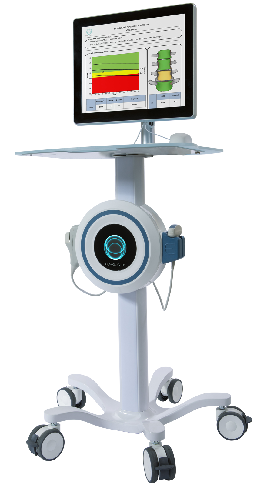
Radiation-Free REMS Technology based scanning for bone quality assessment
Routine osteoporosis screening
Verify response to anti‑osteoporotic medications
Surgical planning
Fracture assessment
REMS vs. DXA at a Glance
REMS Technology:
Radiation Exposure - NONE
Measurement Technique - ULTRASOUND
Bone Quality Assessment - Yes (Fragility Score)
Clinical Use - Emerging
Scan Duration - 40-80 seconds
What is REMS?
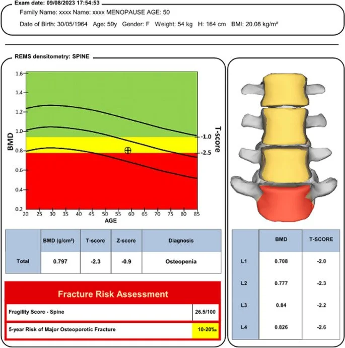
Radiofrequency Echographic Multi Spectrometry (REMS) is a non‑invasive ultrasound technique for evaluating bone health. By analyzing the raw radio‑frequency signals reflected from the lumbar spine and hip, REMS estimates bone‑mineral density (BMD) and reveals micro‑architectural information linked to fracture risk—all without any ionizing radiation.
How the Technology Works
Ultrasound‑based acquisition High‑frequency sound waves are sent through the target bone, and the returning echoes are captured in their unfiltered, raw form.
Advanced signal processing Proprietary algorithms screen out motion and soft‑tissue artifacts, then convert the cleaned signal into quantitative BMD values.
Fragility scoring Beyond density, REMS generates a “Fragility Score” that reflects bone quality, helping clinicians gauge fracture risk more comprehensively.
Old, traditional Dual X-Ray (DXA) Technology:
Radiation Exposure - Low-dose X-ray
Measurement Technique - X-ray
Bone Quality Assessment - No
Clinical Use - Established Gold Standard
Scan Duration - 10-20 minutes
Clinical Advantages
Safer monitoring Zero radiation enables frequent testing—for example, during treatment titration or in younger populations where X‑ray exposure is a concern.
Improved fracture prediction Multiple studies show REMS closely tracks DXA BMD yet identifies high‑risk patients that DXA can miss, particularly when degenerative changes artificially elevate DXA readings.
Workflow efficiency Short scan times and automated analysis support high‑throughput screening in both primary‑care and specialist settings.
Key Use Cases
Routine osteoporosis screening in post‑menopausal women and older men.
Therapy monitoring to verify early response to anti‑osteoporotic medications.
Pre‑operative assessment where bone integrity influences surgical planning.
Secondary osteoporosis evaluation in conditions such as diabetes, chronic kidney disease, or long‑term steroid use.
Takeaway
REMS radiation-free bone quality scanning delivers a fuller picture of bone health—density and quality—without exposing patients to radiation. As evidence mounts and clinical adoption widens, this fast, safe, and insightful technology is poised to redefine osteoporosis diagnosis and fracture‑risk management.